DR ANTHONY MELVIN CRASTO,WorldDrugTracker, helping millions, A 90 % paralysed man in action for you, I am suffering from transverse mylitis and bound to a wheel chair, With death on the horizon, nothing will not stop me except God................DR ANTHONY MELVIN CRASTO Ph.D ( ICT, Mumbai) , INDIA 25Yrs Exp. in the feld of Organic Chemistry,Working for GLENMARK GENERICS at Navi Mumbai, INDIA. Serving chemists around the world. Helping them with websites on Chemistry.Million hits on google, world acclamation from industry, academia, drug authorities for websites, blogs and educational contribution
Showing posts with label NMR spectroscopy. Show all posts

Saturday 2 January 2016

NMR SPECTROSCOPY

Rico positive Umm alkaloid
1 H NMR  

1 it can be seen that from the H NMR, in the compound in the number of protons (integral value) and is dependent on the electronic state chemical shift (ppm) and, dependent on the number of adjacent proton coupling there is. To make a structural analysis, first, one look at the H NMR, where the chemical shift of some, there are any number of protons, and then determines whether you are what coupling (binding constant). Introduction coupling for. Proton of the molecule, as shown above


geminal and vicinal when magnetically non-equivalent proton is present in the position, it makes the coupling (division). Proton each other that the coupling together, the samecoupling constant ( J value) has a (crack width), sp 3 in the coupling of the geminal proton each other is attached to the hybridized carbon is 10 Hz position, vicinal proton each other For, it has a value that depends on the dihedral angle of the two protons as shown in the figure below. When the dihedral angle is close to 0 and 180 ° takes a large value, you can see that taking a small value when it is close to 90 °. 


Here an example of protons that are coupled. 


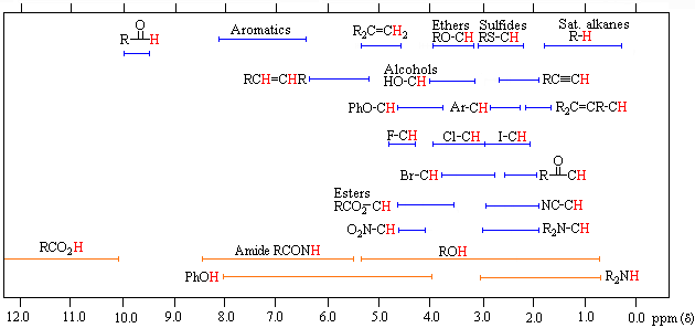
The figure above shows the picture of a case where there proton is a total of two geminal and vicinal proton coupling. ①: a state in which nothing has been coupling. ( s: singlet ) ②: state in which the coupling between one proton coupling constant a. ( D: doublet ) ③: ② state proton is another proton coupling in a more binding constant a of. ( dd: double doublet )     or ① state proton is coupled magnetically equivalent to two protons. ( t: Triplet ) ④: ② state proton is another protons and coupling in the near value (b) to further coupling constant a of. ( dd )     (slightly overlapping the center of the mountain, become larger than the outside of the mountain.) ⑤: ② state proton is another protons and coupling with significantly different value (c) the further coupling constant a of. ( dd ) (the height of the peak of 1: 1: 1: to 1) above diagram depicts the picture of a case where there proton is coupled with a total of three of geminal and vicinal proton. ⑥: ③ state proton is another proton coupling in a more binding constant a of. ( ddd: double double doublet )     or ① state proton is coupled magnetically equivalent to three protons. ( Q: Quartet ) (height of the peak is approximately 1: 3: 3: become 1)⑦: ③ state proton is another proton coupling further in coupling constant a smaller value (d) of. ( ddd ) ⑧: ③ state proton is another protons and coupling with a very large value (e) than further coupling constant a of. ( ddd ) then chemical shift for. The above figure, proton in a variety of environments can be taken, it shows the approximate chemical shift values. Proton bound to a benzene ring or a double bond takes a value of 6 ~ 8 ppm, methine present in the oxygen base of the ester, methylene, you can see that it is 4 ~ 5 ppm. Moreover, methine nitrogen atom is bonded to an oxygen atom, a methylene is 2.6 ~ 4.1 ppm, a hydrocarbon that does not bind to their heteroatom takes the value of 0.8 ~ 2.2 ppm. In addition, the integral value for (number of protons).


In the figure (1) to (5) all is 1 pieces of protons, and chemical shift, it is shown the case where the coupling is different. 
As you can see, even in the same one piece of the proton, we can see that the height of the peak by way of coupling is different. 
Basically, the more to increase the coupling, but peak height tend to be low, the integral value of each proton (area) does not change. 
If the peak height differ greatly even though it has already been a coupling similar, consider the number of protons is different. Well, there is a method of determining the chemical shift, which is one of the center of gravity of the proton is may be obtained a. Singlet: The value of the apex of the peak, doublet: the average value of the vertices of the two peaks triplet: The value of the apex of the peak of the middle quartet: the average value of the apex of the peak of the both ends or ... asked how would each person? By the way, the case of the above figure, (1) In the case of: There is one proton to 2.01 ppm, coupling s (singlet). (2) In the case of: located in one of the protons is 5.42 ppm, coupling d (doublet), coupling constant (J) is          (5.423-5.410) × 800 = 10.4 from 10.4 Hz. (3) In the case of: There is one proton to 3.40, coupling dd ​​(double doublet), coupling constant (J) is          (3.409-3.393) ÷ 2 × 800 = 6.4 than 6.4, 6.4 Hz. (4) In the case of: Yes to the 6.32 ppm 1 single proton, the coupling dd ​​(double doublet), coupling constant (J) is          (6.339-6.318) × 800 = 16.8 than one is 16.8 Hz. The other,          (6.339-6.330) × 800 = 7.2 than 7.2 Hz. In the case of (5): There is one proton to 1.55 ppm, coupling ddd (double double doublet), coupling constant (J) is          (1.563-1.551) ÷ 2 × 800 = 4.8 than 4.8, 4.8 Hz. The other,         (1.563-1.560) × 800 = 2.4 than 2.4 Hz. And it will decipher. Chemical shifts, rounded to two decimal places, on the other hand, the coupling constant is good to round off the second decimal place. ppm of the display 1 when calculating the binding constants from the H NMR should multiplied by the resonant frequency of the device to the difference between the peak apex. In the case of the figure above, it has 800 is used for the calculation formula because using NMR of 800 MHz. Moreover, when put together as data, if the (1), 2.01 (1H, s) In the case of (5), 1.55 (1H, ddd, 4.8, 4.8, 2.4) and will be referred.


13 C NMR

13 it can be seen from C NMR is the number of carbon atoms in the compound is dependent on the electronic state chemical shift (ppm) it is. 
All of the signal is a singlet, you do not need to seek the binding constant. If a large number of protons attached to carbon atoms, or an easily measured, there is a tendency that the height of the signal is high.Chemical shift is good when rounded to 2 decimal places.

From HMQC, C at a distance of Ha and 1 Bond 1 it will give correlation with.
Correlation shown in red, the carbon atoms of 61.9 ppm, represents that the bond is 2.74 and 2.84 ppm proton. 
Moreover, the correlation shown in blue, to a carbon atom of 41.3 ppm, represents that the bond is 2.25 and 2.38 ppm proton. 
Area shown in green, will not overlap a lot of correlation. Analysis give up please. It may be able to be analyzed by other means.
Ha If you pay attention to,
1 H 1 from the H COSY, Ha and Hb, c, obtained correlation with d. 
(Correlation between protons at a distance of 2, 3 bond from Ha) chart While are separated by a triangular red and blue, two regions are symmetrical, that is sufficient to analyze all either one of a pair It will be. For example, correlation and shown in light blue, both the correlation shown in purple, because it represents the correlation of 2.25 ppm and 2.38 ppm proton, and decrypt both, you will duplication of effort. Correlation shown in yellow shows the correlation of 2.56 ppm protons and 1.92 ppm proton. In addition, from the correlation shown in green, of 2.56 ppm proton further shows that there is a correlation with the 2.68 ppm proton.


From HMBC, C is a distance from Ha to 3 Bond 1 , C 2 , C 3 , C 5 , C 6 and the correlation is obtained with.
Correlation shown in red, shows the carbon correlation of 2.56 ppm protons and 39.9 ppm. In addition, the correlation shown in blue, of 2.56 ppm proton, shows that there is a correlation with carbon of 56.1 ppm. Green, correlations shown in purple represents that there is a correlation of two protons 3.00 and 3.06 ppm for the carbon of 52.8 ppm. On the other hand, the correlation shown in yellow shows the correlation between 10.0 ppm of carbon, but also try the attribution of the protons by tracing the correlation, I can not find proton. This correlation is observed in HMQC (1bond), in HMBC, because there is a feature that causes division. The 10.0 ppm proton location where there is a correlation with the carbon of the case will be 0.97 ppm is a midpoint of 0.85 and 1.08 ppm. Such a correlation, when, let's note Ayamarase the analysis. NOESY


From NOESY, Hb in the Ha and spatially close distance, c, d, e, I correlation with i is obtained. NOESY is mainly used when assigning a relative placement. COSY Similarly, blue and red of the area, but has become a nearly symmetrical, etc. If the correlation is poor, who both were firmly analysis would be better. Well, in the NOESY, as observed even COSY, associative even seen a correlation between close proton (2,3bond) It is not a wonder. Therefore, it can be said from the charts NOESY, information excluding the correlation seen in the chart of COSY is especially useful information. Yellow, green, correlations shown in purple shows a NOESY correlation in not observed COSY. With that collect such information in detail, you might be able to infer the relative placement.


Other
Or let's say even with the extended version of the COSY. 
From HOHAHA, Ha and Hb, c, d, e, f, g, h, I correlation is obtained with j. 
(If there are quaternary carbon in the carbon chain, the correlation between the above in the proton can not be obtained basically .Ha and Hk, l, m, n, o, etc..)

Saturday 17 October 2015

NMR Spectroscopy 1: The basics


NMR Spectroscopy 1: The basics

Hello everybody!
This morning, I struggled with NMR spectroscopy. I thought I understand the theory. However, when I was asked to draw a 1H NMR Spectrum of a molecule, I couldn't do it. I wonder what is the problem. So, I thought I put it write here.

Here how it's going to go. Firstly, I am going to write in my own word the theory behind NMR Spectroscopy. Secondly, I am going to confront my problem that is drawing NMR Spectrum of a molecule. Ultimately, I want to make sure that I understand all relevant information and I am able to make use of it for whatever objectives I want to achieve depending on the question.

Before I begin, I would like to share with you the question that I have been asked.

Structure of phenobarbitone. Source: Wikipedia
 Q: Draw the 1H NMR Spectrum you would expect for the drug molecule. Annotate clearly on the spectrum the chemical shift, integration information and splitting patterns you would expect. (5 marks)

OK. Let's begin.

Properties of nuclei.
One of the properties of nuclei is that it has nuclear spin. However, not all nuclei have it. Nuclear spin is quantized and has the symbol I. The exact number of different energy levels that a nucleus has depends on the value of I of that particular isotope. (For my purpose, we do not want to go deeper into this.) Just concentrate on 2 different nuclei that is 13C and 1H. Both have only 2 different energy levels.

NMR uses a strong magnetic field.

In an NMR instrument, there is a large supercooled magnet that provides external magnetic field. When a sample is put into the instrument, all nuclei which possess nuclear spin will be in their lowest energy state. For the case of 13C and 1H, they both have two different energy levels; one is aligned with the external magnetic field (therefore, lower energy level) and the other one is against the external magnetic field (therefore, higher energy level).

The difference between the two energy levels can be measured.

Nuclei that possess nuclear spin and be in their lowest energy state in an external field is said to interact with the magnetic field. The difference between the two energy levels can be measured (how it is done will be explained later). The difference is given as :
ΔE = hf where ΔE is the difference in energy, h is the planck constant (h = 6.62606957 × 10⁻³⁴ m2 kg / s ), and f is the frequency of the radio waves.
ΔE depends on:
  1. How strong the magnetic field is
  2. Magnetic properties of the nucleus itself
NMR uses radio waves to provide energy to flip nuclei from lower energy spin state to higher energy spin state.

Radio wave is in the frequency of 10^4 Hz to 10^8 Hz. The specificity of the spectrum is determined by the frequency of the radio waves i.e. each nuclei require certain frequency of radio waves to flip it from lower energy spin state to higher energy spin state.

The sample is irradiated with short pulse of radio waves of specific frequency. After irradiation, nuclei that is promoted to higher energy spin state will return to lower energy spin state. Energy is released as the nuclei fall back down and this energy can be measured.

Measurement obtained from NMR spectroscopy has to be Fourier transformed to obtained meaningful spectrum.

This is the confusing bit but since I am not an analytical chemist nor physical chemist, we can just accept the fact that the measurement of energy released has to be mathematically treated to produce typical NMR spectrum that we pharmacist can analysed.

The following diagram is an example of a typical NMR spectrum.
1H NMR spectrum of ethanol. Source: Wikipedia
At this moment, we are not going to interpret. I am just showing you the typical NMR spectrum. In reality, NMR spectrum is way more complicated than this and many research lab has been developing algorithm to improve the resolution (that is the ability to see each peak as distinct). We do not want to get deeper into this as this is for advanced study. I don't need it at the moment.

What kind of information does NMR Spectrum gives?
Fundamentally, NMR spectrum enables you to detect atomic nuclei and identify what environment it is in.

Several important information that you can gain from 1H NMR spectrum. (refer to the 1H NMR spectrum of ethanol for clarity)
  1. The number of peaks indicate the number of proton environment.
  2. The area under the peak or integration information tells you how many protons are there. Note: This information is in the form of ratio. In order to correctly identify how many protons are present, other techniques like Mass Spectroscopy should be used to precisely determine number of atoms in the molecule.
  3. The splitting pattern indicates the number of neighbouring nuclei that the nuclei interact with. Note that if the nuclei interact, they will have the same coupling constant. (usually written as J but it is not shown in this spectrum).
  4. Chemical shift tells you about the nature of the environment. This is the most difficult part of interpreting an NMR spectrum. However, we are going to go through this at some point in this post. Just wait for it.
Before we go through information in the NMR spectrum, I would like to first introduce you to chemical shift scale.

Chemical shift scale

The units for abscissa (or x-axis) is parts per million. Why is this the case? This is because the frequency of each nuclei environment depends on the strength of the external magnetic field. If the sample is run on a different NMR instrument or different strength of the magnetic field, the frequency at which that nuclei resonates will be different.
Before I continue, I would like to explain the meaning of the term resonates. In NMR spectroscopy, when nuclei is said to resonate it means that nuclei is absorbing energy from the radio waves so that it can be promoted to higher energy level spin state. As it falls down to lower energy level spin state, it releases energy which through several steps gives the NMR spectrum.
Hence, it will be difficult to say exactly where our signal is. Therefore, we defined our signal position by how far it is from a reference sample (tetramethylsilane is often used as reference), as a fraction of the operating frequency of the instrument.

Since the instrument usually has frequency of up to 900MHz and atomic nuclei resonates at frequency in Hz, it makes sense to use part per million. (Hz in MHz)

The ordinate represents relative amount of nuclei.

What gives each nuclei environment a distinct chemical shift?

Recall that we measure the energy released by nuclei as it falls down from higher energy level spin state to lower energy level spin state.
Each nuclei in a molecule resonate at different frequency. Why is this the case?
Logical deduction that we can make is is that the ΔE is difference for each nuclei. Recall that ΔE = hf.

Though the external magnetic field is constant for all nuclei, the magnetic field experienced by each nuclei is not exactly the same. .
Each nucleus is surrounded by electrons. In a magnetic field, electrons can generate tiny electrical current. Consequently, this electrical current will generate magnetic field.
Basic physics guys. Just to remind myself again. Suppose we have a wire and we coil it so that it forms a solenoid. When we pass current through it, magnetic field is generated.
This magnetic field which we will call local magnetic field will oppose the external magnetic field. Electron distribution around the nucleus vary for each environment. Two possible situations: shielding and deshielding.

When nucleus is protected by electrons around it, it is called shielding as the electrons shield the nucleus from external magnetic field. Consequently, the resonating frequency will shift upfield.
The vice versa is true. (More on this later.)

Take ethanol for example. I suggest you draw the structure in a piece of paper and follow my explanation. Oxygen atom is an electronegative atom which means in ethanol molecule electrons density will be higher towards the oxygen atom. As a result, proton attached to carbon next to oxygen atom will have less electron density hence deshielded. The nuclei will experience more external magnetic field i.e. greater energy difference. From ΔE = hf, we can infer that resonating frequency will also increase. Hence, the signal for CH2 is further downfield with regards to CH2 signal position without the oxygen atom.

I know things has become more difficult. Don't give up. You just need to get your head around it. I suggest you ensure that you understand everything up till this point. Read back from the beginning if necessary.

OK. Are you ready to continue?

Describing chemical shift.

If you find it difficult to follow the previous concept, I think one of the reason is that I haven't introduced the terminology to describe chemical shift.

First, I want to define chemical shift. Chemical shift is the resonating frequency with respect to standard as a fraction of frequency of instrument.

It's hard to explain the terminology without using a diagram. However, if you look into textbook of organic chemistry, you will find one.

When we are saying about the field i.e. magnetic field, we use downfield and upfield.
When we are saying about chemical shift, we use large or small.
When we are saying about frequency, we use high or low.
When we are saying about shielding, we use deshielded or shielded.

I think the best that I can do now is to give you example so that you understand.

So far, I would like to wrap up a few things before we continue.
Why each nuclei resonates at different frequency?

Each nuclei experience different magnetic field due to distribution of electrons around the nuclei leading to either shielding or deshielding.

What happens to resonating frequency when the nuclei is deshielded and vice versa?

Think about it this way. When a nucleus is deshielded, it experiences greater external magnetic field. Consequently, ΔE will increase. According to equation ΔE = hf, as ΔE increases, resonating frequency will also increase. Hence, the position of the signal will be further downfield.

OK. I think we are now ready to go to deeper.

Regions of the 1H NMR spectrum.

Many observations have been made and it is important to memorise the common proton environment.

Proton in:

CH3 = 1 ppm
CH2 = 2 ppm
CH = 3 ppm
Benzene = 7.2 ppm

Also, a diagram to help you remember the rough position of signal.

Our discussion on NMR spectroscopy has not finished. I am going to continue more on it tomorrow.

Wednesday 26 August 2015

Rapid structure determination of microgram-level drug metabolites using HPLC-MS, fraction collection and NMR spectroscopy

.


A robust method for in vitro metabolite generation and facile sample preparation on analytical HPLC was established for rapid structure determination of microgram-level drug metabolites by using high-field NMR equipped with a cryoprobe. A single 1–5 mL incubation of drug candidate (10–30 μM) in microsomes, hepatocytes, or recombinant drug-metabolising enzymes, typically cytochrome P450s and UDP-glucuronosyltransferases, was used for metabolite formation. Following precipitation of proteins and solvent removal, metabolite mixtures were chromatographed with 5–10 injections onto an HPLC-MS system. Metabolites were collected into a 96-well plate, dried, and reconstituted in deuterated NMR solvents. NMR spectra of isolated metabolites were acquired on a 500 MHz spectrometer equipped with a 5 mm cryogenic probe. The methodology has been successfully employed as an extension of HPLC-MS/MS-based metabolite identification and applied frequently to 0.5–10 μg quantities of metabolite. Most structure determinations were achieved rapidly by 1D 1H NMR with satisfactory signal-to-noise ratios, whereas some required 2D NMR data analysis. This report describes the method development and metabolite structure determination using the model compound trazodone. In addition to trazodone, a large number of examples from our laboratories have proven that the microgram-level NMR method avoids time-consuming preparative-scale metabolite generation and purification and circumvents technical complications associated with online LC-NMR. Most importantly, the turnaround time of metabolite structure determination for metabolically unstable compounds using the present methodology is more in sync with the cycle time during which medicinal chemists modify metabolic softspots while performing other iterative lead optimisation activities, demonstrating a real impact on the drug-discovery process.


Graphical abstract: Rapid structure determination of microgram-level drug metabolites using HPLC-MS, fraction collection and NMR spectroscopy


Rapid structure determination of microgram-level drug metabolites using HPLC-MS, fraction collection and NMR spectroscopy

*Corresponding authors
aPharmaceutical Candidate Optimization, Bristol-Myers Squibb Research & Development, 5 Research Parkway, Wallingford, USA
E-mail: yuezhong.shu@bms.com
Anal. Methods, 2010,2, 1542-1549

DOI: 10.1039/C0AY00290A
 http://pubs.rsc.org/en/content/articlelanding/2010/ay/c0ay00290a/unauth#!divAbstract


///////

Saturday 14 June 2014

NMR Spectroscopy of Stereoisomers


Introduction

Nuclear magnetic resonance (NMR) spectroscopy is a very useful tool used widely in modern organic chemistry. It exploits the differences in the magnetic properties of different nuclei in a molecule to yield information about the chemical environment of the nuclei, and subsequently the molecule, in question. NMR analysis lends itself to scientists more easily than say the more cryptic data achieved form ultraviolet or infared spectra because the differences in magnetic properties lend themselves to scientists very well. The chemical shifts that are characteristic of different chemical environments and the multiplicity of the peaks fit well with our conception of the way molecules are structured.
Using NMR spectroscopy, we can differentiate between constitutional isomers, stereoisomers, and enantiomers. The later two of these three classifications require close examination of the differences in NMR spectra associated with changes in chemical environment due to symmetry differences; however, the differentiation of constitutional isomers can be easily obtained.

Constitutional isomerism

Nuclei both posses charge and spin, or angular momentum, and from basic physics we know that a spinning charge generates a magnetic moment. The specific nature of this magnetic moment is the main concern of NMR spectroscopy.
For proton NMR, the local chemical environment makes different protons in a molecule resonate at different frequencies. This difference in resonance frequencies can be converted into a chemical shift (δ) for each nucleus being studied. Because each chemical environment results in a different chemical shift, one can easily assign peaks in the NMR data to specific functional groups based upon president. Presidents for chemical shifts can be found in any number of basic NMR text. For example, Figure 1 shows the spectra of ethyl formate and benzyl acetate. In the lower spectra, benzyl acetate, notice peaks at δ = 1.3, 4.2, and 8.0 ppm characteristic of the primary, secondary, and aromatic protons, respectively, present in the molecule. In the spectra of ethyl formate (Figure 1b), notice that the number of peaks is is the same as that of benzyl acetate (Figure 1a); however, the multiplicity of peaks and their shifts is very different.
Figure 1: 1H NMR spectra of (a) ethyl formate and (b) benzyl acetate.
Figure 1 (graphics1c.jpg)
The difference between these two spectra is due to geminal spin-spin coupling. Spin-spin coupling is the result of magnetic interaction between individual protons transmitted by the bonding electrons between the protons. This spin-spin coupling results in the speak splitting we see in the NMR data. One of the benefits of NMR spectroscopy is the sensitivity to very slight changes in chemical environment.

Stereoisomerism

Diastereomers

Based on their definition, diastereomers are stereoisomers that are not mirror images of each other and are not superimposable. In general, diastereomers have differing reactivity and physical properties. One common example is the difference between threose and erythrose (Figure 2).
Figure 2: The structures of threose and erythrose.
Figure 2 (FigNMR1.jpg)
As one can see from Figure 2, these chemicals are very similar each having the empirical formula of C4H7O4. One may wonder: how are these slight differences in chemical structure represented in NMR? To answer this question, we must look at the Newman projections for a molecule of the general structure (Figure 3).
Figure 3: Newman projections of a general diastereomer.
Figure 3 (graphics3.jpg)
One can easily notice that the two protons represented are always located in different chemical environments. This is true because the R group makes the proton resonance frequencies v1(I) ≠ v2(III), v2(I) ≠ v1(II), and v2(II) ≠ v1(III). Thus, diastereomers have different vicinal proton-proton couplings and the resulting chemical shifts can be used to identify the isomeric makeup of the sample.

Enantiomers

Enantiomers are compounds with a chiral center. In other words, they are non-superimposable mirror images. Unlike diastereomers, the only difference between enantiomers is their interaction with polarized light. Unfortunately, this indistinguishability of racemates includes NMR spectra. Thus, in order to differentiate between enantiomers, we must make use of an optically active solvent also called a chiral derivatizing agent (CDA). The first CDA was (α-methoxy-α-(trifluoromethyl)phenylacetic acid) (MTPA also known as Mosher's acid) (Figure 4).
Figure 4: The structure of the S-isomer of Mosher's Acid (S-MTPA)
Figure 4 (FigNMR2.jpg)
Now, many CDAs exist and are readily available. It should also be noted that CDA development is a current area of active research. In simple terms, one can think of the CDA turning an enantiomeric mixture into a mixture of diastereomeric complexes, producing doublets where each half of the doublet corresponds to each diastereomer, which we already know how to analyze. The resultant peak splitting in the NMR spectra due to diastereomeric interaction can easily determine optical purity. In order to do this, one may simply integrate the peaks corresponding to the different enantiomers thus yielding optical purity of incompletely resolved racemates. One thing of note when performing this experiment is that this interaction between the enantiomeric compounds and the solvent, and thus the magnitude of the splitting, depends upon the asymmetry or chirality of the solvent, the intermolecular interaction between the compound and the solvent, and thus the temperature. Thus, it is helpful to compare the spectra of the enantiomer-CDA mixture with that of the pure enantiomer so that changes in chemical shift can be easily noted.

Bibliography

  • H. Günther, NMR Spectroscopy: Basic Principles, Concepts, and Applications in Chemistry, John Wiley & Sons, Chichester (1996).
  • F. A. Bovey, Nuclear Magnetic Resonance Spectroscopy, 2nd Ed, Academic, New York (1988).
  • S. Braun, H.-O. Kalinowski, S. Berger, 100 and More Basic NMR Experiments: A Practical Course, VCH, Weinheim (1996).
  • A. E. Derome, Modern NMR Techniques for Chemistry Research , Pergamon, Oxford (1987).
  • J. A. Dale and H. S. Mosher, J. Am. Chem. Soc., 1973, 95, 512.

King, S.; Barron, A. NMR Spectroscopy of Stereoisomers, OpenStax-CNX Web site. http://cnx.org/content/m38355/1.1/, May 16, 2011.

Wednesday 4 December 2013

DOXOFYLLINE SPECTRAL DATA

DOXOFYLLINE
69975-86-6  CAS NO
7-(1,3-dioxolan-2-ylmethyl)-1,3-dimethylpurine-2,6-dione
FormulaC11H14N4O4 
Mol. mass266.25 g/mol
Doxofylline (INN), (also known as doxophylline) is a xanthine derivative drug used in the treatment of asthma.[1]
It has antitussive and bronchodilator[2] effects, and acts as aphosphodiesterase inhibitor.[3]
In animal and human studies, it has shown similar efficacy to theophylline but with significantly fewer side effects.[4]
Unlike other xanthines, doxofylline lacks any significant affinity for adenosine receptorsand does not produce stimulant effects. This suggests that its antiasthmatic effects are mediated by another mechanism, perhaps its actions on phosphodiesterase.[1]

Doxofylline, [7-(1, 3-dioxolan-2-ylmethyl)-3, 7-dihydro-1, 3-dimethyl-1H-purine-2, 6-dione] is a new bronchodilator xanthine based drug which differs from theophylline by the presence of dioxalane group at position 7. It is used in the treatment of bronchial asthma, chronic obstructive pulmonary disease (COPD), and chronic bronchitis . The mechanism of action is similar to that of theophylline in that it inhibits phosphodiesterase (PDE-IV), thereby preventing breakdown of cyclic adenosine monophosphate (cAMP). Increase in cAMP inhibits activation of inflammatory cells resulting in bronchodilating effect [52]. In contrast to theophylline, doxofylline has very low affinity towards adenosine A1 and A2 receptors which explain its better safety profile
Doxofylline (7-(l,3-dioxalan-2-ylmethyl)-theophylline) is a drug derived from theophylline which is used in therapy as a bronchodilator, with anti-inflammatory action, in reversible airway obstruction. It is commonly administered in doses ranging from 800 to 1200 mg per day, orally, according to a dosage which provides for the intake of two to three dosage units per day in order to maintain therapeutically effective haematic levels. The doxofylline tablets commercially available generally contain 400 mg of active ingredient and release almost all the drug within one hour from intake. The half- life of the drug is around 6-7 hours and for this reason several administrations are required during the 24-hour period.
Obviously a drop in haematic concentration of the drug in an asthmatic patient or patient suffering from COPD (chronic obstructive pulmonary disease) can result in serious consequences, in which case the patient must have recourse to rescue medication, such as salbutamol inhalers.
Pharmaceutical techniques for obtaining the modified release of drugs have been known for some time, but no modified release formulation of doxofylline is known. In fact the present inventors have observed that there are significant difficulties in the production of a doxofylline formula that can be administered only once a day and in particular have encountered problems correlated with bioequivalence.
Various attempts to formulate doxofylline in modified release systems, with different known polymers, have not provided the desired results, i.e. a composition that can be administered once a day, bio equivalent to the plasmatic concentration obtained with the traditional compositions currently on sale. In fact currently, dosage units containing 400 mg of active ingredient are currently administered two/three times a day for a daily average of approximately 1000 mg of active ingredient, a dosage considered necessary to maintain the therapeutic haematic levels of doxofylline.
Such a dosage unit is currently marketed by Dr. Reddy's Laboratories Ltd as DOXOBID and has the following quali-quantitative composition: doxofylline (400 mg), colloidal silicon dioxide (13 mg), corn starch (63 mg), mannitol (40 mg), povidone (7 mg), microcrystalline cellulose (64 mg), talc (30 mg), magnesium stearate (3 mg) and water (0.08 ml).
  1.  Cirillo R, Barone D, Franzone JS (1988). "Doxofylline, an antiasthmatic drug lacking affinity for adenosine receptors". Arch Int Pharmacodyn Ther 295: 221–37.PMID 3245738.
  2. Poggi R, Brandolese R, Bernasconi M, Manzin E, Rossi A (October 1989). "Doxofylline and respiratory mechanics. Short-term effects in mechanically ventilated patients with airflow obstruction and respiratory failure"Chest 96 (4): 772–8.doi:10.1378/chest.96.4.772PMID 2791671.
  3.  Dini FL, Cogo R (2001). "Doxofylline: a new generation xanthine bronchodilator devoid of major cardiovascular adverse effects". Curr Med Res Opin 16 (4): 258–68.doi:10.1185/030079901750120196PMID 11268710.
  4. Sankar J, Lodha R, Kabra SK (March 2008). "Doxofylline: The next generation methylxanthine". Indian J Pediatr 75 (3): 251–4. doi:10.1007/s12098-008-0054-1.PMID 18376093.
  • Dali Shukla, Subhashis Chakraborty, Sanjay Singh & Brahmeshwar Mishra. Doxofylline: a promising methylxanthine derivative for the treatment of asthma and chronic obstructive pulmonary disease. Expert Opinion on Pharmacotherapy. 2009; 10(14): 2343-2356, DOI 10.1517/14656560903200667, PMID 19678793
At present, domestic synthetic Doxofylline composed of two main methods: one is by the condensation of theophylline prepared from acetaldehyde and ethylene glycol, but this method is more complex synthesis of acetaldehyde theophylline, require high periodate oxidation operation. Another is a halogenated acetaldehyde theophylline and ethylene glycol is prepared by reaction of an organic solvent, the method were carried out in an organic solvent, whereby the product Theophylline caused some pollution, conducive to patients taking.
current domestic Doxofylline synthetic methods reported in the literature are: 1, CN Application No. 94113971.9, the name "synthetic drugs Doxofyllinemethod" patents, the patent is determined by theophylline with a 2 - (halomethyl) -1,3 - dimethoxy-dioxolane in a polar solvent, with a base made acid absorbent,Doxofylline reaction step. 2,  CN Application No. 97100911.2, entitled "Synthesis of Theophylline," the patent, the patent is obtained from 7 - (2,2 - dialkoxy-ethyl) theophylline with ethylene glycol in N, N-dimethylformamide solvent with an alkali metal carbonate to make the condensing agent, p-toluenesulfonic acid catalyst in the condensation Doxofylline.
Doxofylline of xanthine asthma drugs, and its scientific name is 7 - (1,3 - dioxolan - ethyl methyl) -3,7 - dihydro-1,3 - dimethyl-1H - purine-2 ,6 - dione. The drug developed by the Italian Roberts & Co. in 1988, listed its tablet tradename Ansimar. This product is compared with similar asthma drugs, high efficacy, low toxicity, oral LD50 in mice is 1.5 times aminophylline, non-addictive. Adenosine and its non-blocking agents, it does not produce bronchial pulmonary side effects, no aminophylline like central and cardiovascular system. U.S. patent (US4187308) reported the synthesis of doxofylline, theophylline and acetaldehyde from ethylene glycol p-toluenesulfonic acid catalyst in the reaction of benzene as a solvent Doxofylline. Theophylline acetaldehyde by the method dyphylline derived reaction with a peroxy periodate or 7 - (2,2 - dialkoxy-ethyl) ammonium chloride aqueous solution in the decomposition of theophylline converted to acetaldehyde theophylline . Former method is relatively complex, and the high cost of using periodic acid peroxide, low yield after France. And theophylline acetaldehyde and ethylene glycol solvent used in the reaction of benzene toxicity, harm to health, and the yield is low, with an average around 70%, not suitable for industrial production.
Theophylline-7-acetaldehyde (I) could react with ethylene glycol (II) in the presence of p-toluenesulfonic acid in refluxing benzene to produce Doxofylline.
.........................
the reaction is:
Figure CN102936248AD00041
a, anhydrous theophylline and bromoacetaldehyde ethylene glycol as the basic raw material, purified water as a solvent with anhydrous sodium carbonate as acid-binding agent;
UV (95% C2H5OH, nm) λmax273 (ε9230); λmin244 (ε2190)
IR (KBr, cm-1) 1134 (CO); 1233 (CN) ; 1547 (C = N); 1656 (C = C); 1700 (C = O); 2993 (CH)
1H-NMR [CDCl3, δ (ppm)] 3.399 (s, 3H, N-CH3); 3.586 (S, 3H, N-CH3); 3.815-3.885 (m, 4H, OCH2 × 2); 4.581 (d, 2H, CH2); 5.211 (t, 1H, CH ); 7.652 (S, 1H, CH = N)
13C-NMR [CDCL3, δ (ppm)] 27.88 (CH3); 29.69 (CH3); 47.87 (CH2); 65.37 ( OCH2); 100.76 (CH); 107.26 (C = C); 142.16 (CH = N); 148.22 (C = C); 151.59 (C = O); 155.25 ( C
.........................
Spectral data of doxofylline
The ESI mass spectrum exhibited a protonated molecular ion peak at m/z 267 in positive ion mode indicating the molecular weight of 266. The tandem mass spectrum showed the fragment ions m/z 223, 181.2, 166.2, 138.1, 124.1 and 87.1.
Inline image 2
Inline image 5
Inline image 6
The FT-IR spectrum, two strong peaks at 1697cm-1 and 1658cm-1 indicated presence of two carbonyl groups. A strong peak at frequency 1546cm-1 indicated presence of C=N stretch. A medium peak at 1232cm-1 was due to C-O stretch
Inline image 3
FT IR
1H and 13C-NMR spectra of doxofylline and its degradation products were recorded by using Bruker NMR 300MHz instrument with a dual broad band probe and z-axis gradients. Spectra were recorded using DMSO-d6 as a solvent and tetramethylsilane as an internal standard.

Inline image 1
1H NMR
Inline image 4
13 C NMR
COMPARISONS
Inline image 9
Inline image 8
Inline image 7